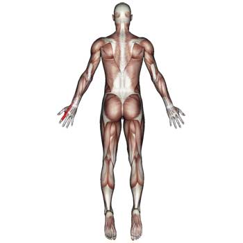
Palmaris Longus Muscle Pain

The palmaris longus muscle contributes to pain in the wrist, the palm of the hand, and will sometimes extend up into the forearm.
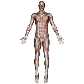
The palmaris longus muscle contributes to pain in the wrist, the palm of the hand, and will sometimes extend up into the forearm.

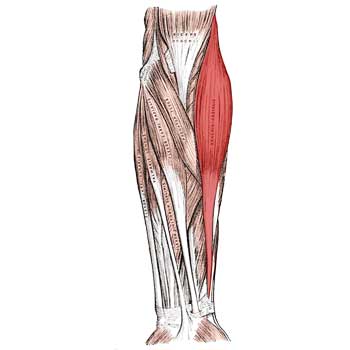
Muscle anatomy of the palmaris longus includes origin, insertion, action, innervation and vascular supply. Actions include agonists and antagonists for each movement.

Muscle anatomy of the flexor carpi ulnaris includes origin, insertion, action, innervation and vascular supply. Actions include agonists and antagonists for each movement.

The flexor carpi ulnaris muscle is located on the inside (pinky side) of the forearm. It contributes to pain in the wrist, palm of the hand, and the ring and little fingers. Pain is sometimes felt in the elbow.

The flexor carpi radialis muscle is located in the front of the forearm. It contributes to pain in the wrist, bottom of the palm that often extends to the thumb pad and thumb. Twisting the wrist and gripping objects is painful.

Muscle anatomy of the pronator quadratus includes origin, insertion, action, innervation and vascular supply. Actions include agonists and antagonists for each movement.

Muscle anatomy of the flexor carpi radialis includes origin, insertion, action, innervation and vascular supply. Actions include agonists and antagonists for each movement.
The extensor indicis muscle is located in the back of the forearm. It contributes to pain in the back of the wrist, hand, and index finger. The pain will often feel like you have sprained or strained the back of the hand. It can also cause charley horse like cramps in the index finger.

Muscle anatomy of the extensor indicis includes origin, insertion, action, innervation and vascular supply. Actions include agonists and antagonists for each movement.

Muscle anatomy of the flexor pollicis longus includes origin, insertion, action, innervation and vascular supply. Actions include agonists and antagonists for each movement.
The flexor pollicis longus muscle is located in the forearm, same side as the thumb. It contributes to pain in the middle joint and tip of the thumb. Pinching motions between the forefinger and thumb can cause intense pain. It can also cause the middle thumb joint to pop and sometimes lock.

Muscle anatomy of the triceps brachii includes origin, insertion, action, innervation and vascular supply. Actions include agonists and antagonists for each movement.

Muscle anatomy of the biceps brachii includes origin, insertion, action, innervation and vascular supply. Actions include agonists and antagonists for each movement.

Muscle anatomy of the coracobrachialis includes origin, insertion, action, innervation and vascular supply. Actions include agonists and antagonists for each movement.
Muscle anatomy of the brachioradialis includes origin, insertion, action, innervation and vascular supply. Actions include agonists and antagonists for each movement.

Muscle anatomy of the brachialis includes origin, insertion, action, innervation and vascular supply. Actions include agonist and antagonist for each movement.rvation and vascular supply. Actions include agonists and antagonists for each movement.

Muscle anatomy of the supinator includes origin, insertion, action, innervation and vascular supply. Actions include agonists and antagonists for each movement.

Muscle anatomy of the anconeus includes origin, insertion, action, innervation and vascular supply. Actions include agonist and antagonist for each movement.

Muscle anatomy of the extensor carpi radialis longus includes origin, insertion, action, innervation and vascular supply. Actions include agonists and antagonists for each movement
Muscle anatomy of the extensor carpi radialis longus includes origin, insertion, action, innervation and vascular supply. Actions include agonists and antagonists for each movement

Muscle anatomy of the extensor carpi ulnaris includes origin, insertion, action, innervation and vascular supply. Actions include agonists and antagonists for each movement.

Muscle anatomy of the pronator teres includes origin, insertion, action, innervation and vascular supply. Actions include agonists and antagonists for each movement.