Flexor Hallucis Longus Muscle Anatomy: Origin, Insertion, Action

Flexor hallucis longus muscle anatomy includes origin, insertion, action, innervation and vascular supply. Actions include agonists and antagonists for each movement.
Flexor hallucis longus muscle anatomy includes origin, insertion, action, innervation and vascular supply. Actions include agonists and antagonists for each movement.
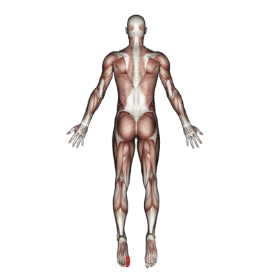
The flexor hallucis longus can cause and contribute to pain in the big toe and ball of the foot. A classic sign of flexor hallucis longus dysfunction is numbness on the bottom of the big toe.
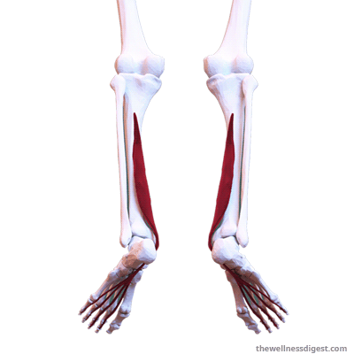
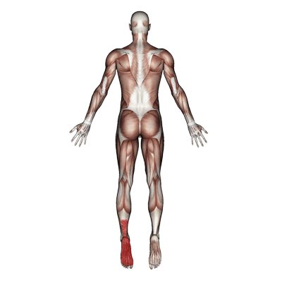
Flexor digitorum longus muscle anatomy includes origin, insertion, action, innervation and vascular supply. Actions include agonists and antagonists for each movement.

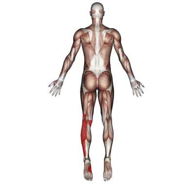
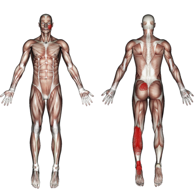
Tibialis posterior muscle anatomy includes origin, insertion, action, innervation and vascular supply. Actions include agonists and antagonists for each movement.

Gastrocnemius muscle anatomy includes origin, insertion, action, innervation and vascular supply. Actions include agonists and antagonists for each movement.

Muscle anatomy of the soleus includes origin, insertion, action, innervation and vascular supply. Actions include agonists and antagonists for each movement.

The flexor digitorum longus muscle contributes to pain in the foot and lower leg. It plays a role in foot cramps, hammertoes, and claw toes.
The tibialis posterior muscle contributes to pain in the lower leg above the heel. This pain will often descend into the heel and the bottom of the foot. The pain may radiate into the lower calf. Dysfunction in muscle contributes to fallen arches and weak ankles that collapse inward.
The gastrocnemius muscle contributes to pain in the back of the knee, lower leg, ankle, and foot arch pain. The muscle is known to contribute to lower leg cramps.
Edit Snippet
The soleus muscle can cause and contribute to pain in the heel, ankle, and back of the knee. It can also cause pain in the low back on the same side of the affected leg. Dysfunction of the soleus muscle can also contribute to swelling in the foot and ankle. Occasionally a trigger point at the bottom and outside of the muscle can contribute to pain in the jaw and side of the head.

The semispinalis cervicis is a major contributor to headaches, especially those in which the pain concentrates at the base of the skull extending up the back of the head. It can also contribute to tingling and burning in the back of the head and scalp.

The adductor magnus muscle can cause groin, pelvic, and thigh pain. The pain ranges from dull annoying aches to sharp, stabbing pain.

Muscle anatomy of the rectus femoris includes origin, insertion, action, innervation and vascular supply. Actions include agonists and antagonists for each movement.

The tensor fasciae latae muscle is located toward the front of the hip. The muscle contributes to hip pain and stiffness. It makes straightening the thigh at the hip difficult and painful.

Muscle anatomy of the buccinator includes origin, insertion, action, innervation and vascular supply. Actions include agonists and antagonists for each movement.

The buccinator muscle is located in the cheek. It can contribute to pain in the cheek, upper teeth and mouth. The pain in the upper teeth often feels like an abscessed tooth. It can also cause pain while chewing and difficulty swallowing.

Muscle anatomy of the extensor digitorum includes origin, insertion, action, innervation and vascular supply. Actions include agonists and antagonists for each movement.

The extensor digitorum is located in the back of the forearm. It contributes to pain in the back of the hand and middle finger which sometimes radiates up into the back of the wrist and the back of the forearm. Pain is occasionally felt in the front of the wrist, just below the palm.

The flexor digitorum profundus muscle is found in the front of the forearm. It can contribute to pain and twitching in the four fingers. It can affect one or any combination of the fingers. It can also cause trigger finger, where a finger will lock in a bent position.

Muscle anatomy of flexor digitorum profundus includes origin, insertion, action, innervation and vascular supply. Actions include agonists and antagonists for each movement.

The flexor digitorum superficialis is located in the front of the forearm. It contributes to pain in the four fingers, the palm, and occasionally the wrist. The muscle is a major contributor to trigger finger.

Muscle anatomy of the flexor digitorum superficialis includes origin, insertion, action, innervation and vascular supply. Actions include agonists and antagonists for each movement.