Lumbrical Muscles Hand Origin, Insertion, Action

Muscle anatomy of the lumbricals muscles of the hand includes origin, insertion, action, innervation and vascular supply. Actions include agonists and antagonists for each movement.
Muscle anatomy of the lumbricals muscles of the hand includes origin, insertion, action, innervation and vascular supply. Actions include agonists and antagonists for each movement.
The adductor pollicis muscle contributes to pain in the thumb and thumb pad.

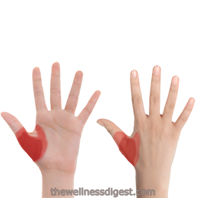
The lumbrical muscles of the hand contribute to pain in the back of the hand and the fingers. Pain in the index finger and the little finger are the most common. Stiffness in the finger joints mimics arthritis pain. Stiffness and pain when opening and closing the hand is also common.

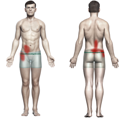
The quadratus lumborum muscle is known for sharp pain in the lower back and aching hip pain. Contributes to pain in the buttocks, groin and abdominal areas.

Quadratus lumborum origin, insertion, action, innervation. Agonists and antagonists are listed for each muscle action.
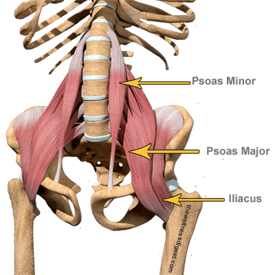
Muscle anatomy of the iliopsoas muscle group includes origin, insertion, action, innervation and vascular supply. Actions include agonists and antagonists for each movement.
The iliopsoas muscles can contribute to lower back, abdomen, groin, upper leg, and pelvic pain. Standing from a sitting position is painful and an indication of iliopsoas dysfunction.

Muscle anatomy of the popliteus muscle includes origin, insertion, action, innervation and vascular supply. Actions include agonists and antagonists for each movement.

Anatomy of the oblique muscles includes origin, insertion, action, innervation and vascular supply. Actions include agonists and antagonists for each movement.

The oblique muscles contribute to pain on your side in the rib cage and waist area, lower abdomen, groin, and pelvis. They can also contribute to heartburn, indigestion, bladder pain and incontinence, and testicle pain.

The rectus abdominis contributes to pain in the area around the sternum, across the upper back, across the buttocks, the lower abdomen, the pelvis. and testicles. It can also contribute to heartburn, indigestion, feeling bloated as well as mimicking urinary tract infection pain.

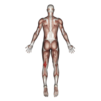
The popliteus muscle contributes to pain and stiffness behind the knee. The muscle is particularly affected by PCL injury and repair surgery.

Muscle anatomy of the rectus abdominis muscle includes origin, insertion, action, innervation and vascular supply. Actions include agonists and antagonists for each movement.

Muscle anatomy of the plantaris muscle includes origin, insertion, action, innervation and vascular supply. Actions include agonists and antagonists for each movement.
The plantaris muscle is a very small muscle located behind the knee. It contributes to pain behind the knee and is occasionally involved in cramps in the calf.

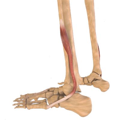
The peroneus tertius contributes to pain on at the top of the foot, ankle and heel. Pain greatly increases when walking and jogging.

Muscle anatomy of the peroneus tertius includes origin, insertion, action, innervation and vascular supply. Actions include agonists and antagonists for each movement.

Muscle anatomy of the peroneus longus includes origin, insertion, action, innervation and vascular supply. Actions include agonists and antagonists for each movement.

The peroneus longus and peroneus brevis contribute to pain on the outside of the ankle and foot. The muscles are contributors to weak ankles and foot drop.
Muscle anatomy of the peroneus brevis includes origin, insertion, action, innervation and vascular supply. Actions include agonists and antagonists for each movement.

The tibialis anterior muscle can cause pain in the big toe, the ankle, and shin. It also contributes to a weak ankle, shin splints, and foot drop.

Extensor digitorum longus muscle anatomy includes origin, insertion, action, innervation and vascular supply. Actions include agonists and antagonists for each movement.